

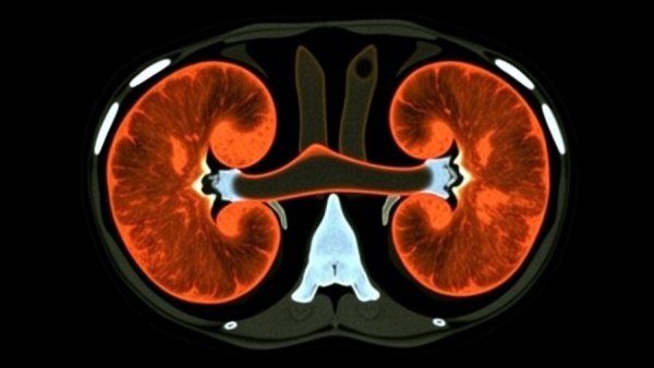
Understanding Supernumerary Kidneys: A Rare Discovery
In a remarkable medical incident, a 58-year-old man in India experienced abdominal pain, jaundice, and loss of appetite, which led him to seek medical help. Initially diagnosed with periampullary adenocarcinoma — a form of cancer near where the bile and pancreatic ducts meet — the man underwent a CT scan to stage his cancer before a planned surgery. However, what radiologists uncovered was nothing short of extraordinary: not just one, but two fused kidneys on his right side, merged together in an S-shaped configuration.
This peculiar discovery was met with intrigue and concern. Radiologists Shreya Shukla, Tushar Sirohi, and Satyendra Narayan Singh published their findings in Radiology Case Reports, shedding light on an anatomical anomaly that surprisingly affects fewer than 100 individuals documented globally. Both kidneys possessed their distinct arteries and urine-collecting systems, functioning independently but linked together in a way that complicates their classification.
The Complex Anatomy of Fused Kidneys
Radiological analysis revealed that the upper kidney measured 7.6 by 3.8 centimeters while the lower kidney measured 7 by 3 centimeters, with the man’s left kidney being normal in size and position. The upper kidney had a typical orientation, but the lower kidney was abnormally rotated. This unusual anatomy raised critical concerns, particularly as the patient prepared for a pancreaticoduodenectomy, known colloquially as the Whipple procedure.
Surgeons needed to pay close attention to the unique arterial and urinary drainage structures associated with the fused kidneys. The lower positioning of the kidney arteries introduced the risk for surgical complications during the procedure, especially within the retroperitoneal space, where both kidneys typically reside. Such risks are not isolated, as documented cases have demonstrated similar complications in other patients with supernumerary kidneys.
The Surgical Implications of Kidney Anomalies
Understanding the nuances of these anatomical variations is vital for healthcare professionals. The authors of the mentioned case stressed the importance of detailed CT mapping before surgery, something that can greatly influence surgical strategy and patient outcomes. Given the intricate blood supply and drainage systems, careful planning and visualization are paramount in mitigating potential risks during what is already a complex procedure.
The patient underwent surgery without complications, and pathology results indicated a successfully excised cancer with clear margins, suggesting no remaining cancerous cells. This positive outcome reflects the potential for successful interventions, even in the presence of significant anatomical challenges.
What This Case Teaches Us About Kidney Health
The man’s case serves as a pivotal reminder of the complexities of human anatomy and the potential for unexpected findings in medical imaging. Although it is uncommon, being aware of supernumerary kidneys is essential for both patients and healthcare providers to ensure comprehensive care. It highlights the need for thorough medical evaluations that extend beyond initial diagnoses, opening avenues for improved patient care and outcomes.
With the right insights and attention to detail, medical professionals can navigate these complexities and provide effective treatment for patients in unique anatomical situations. This case is an exciting blend of curiosity and clinical significance, showing how unexpected discoveries may lead to better understanding and management of renal health.





Write A Comment